
Raman spectroscopy, a vibrational spectroscopic technique which gives information about molecular structure with a high level of specificity, is begging to be recognized as a high potential technique for the non invasive study of biological samples, in particular on human tissues. In dermatological research it can be used as an efficient tool for examination of skin’s biochemical structure and composition, aspects of interest for diagnosis and dermal application of pharmacological and cosmetic agents. Raman spectroscopy has already been demonstrated to potentially provide an accurate early diagnosis by distinguishing in-vivo different type of skin cancers (1,2) and for detecting of filaggrin-related atopic dermatitis(3).
In our lab, thanks to a wide experience on human skin biology and morphology, we are studying the Raman spectra of ex-vivo human skin, to create a spectral database to index vibration peaks and bands of the skin layers from which we can obtain information regarding their molecular composition, thus potentially highlighting alterations linked to a specific disease (e.g. skin cancer). This project is granted by the Italian Ministry of Health (Ricerca Corrente 2013-2014).
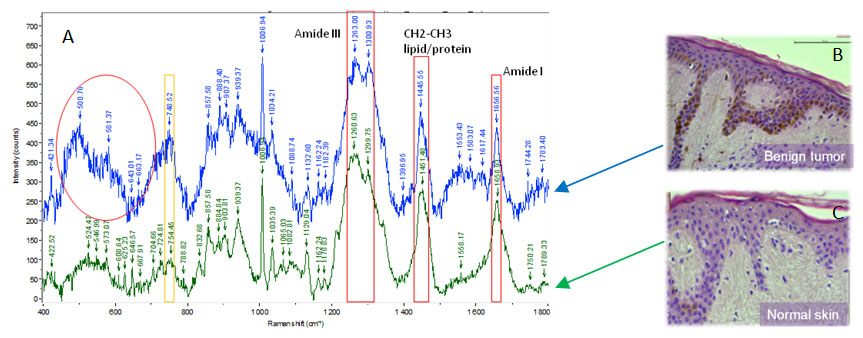
Our results suggest that Raman spectroscopy is able to identify in a specific way the skin chemical composition and its alterations of normal skin vs. tumoral skin. Here below an example of Raman spectra on healthy and benign tumor human skin.
References:
- Lui H et al., Cancer Res 2012;15,72(10):2491-500
- Nijssen A et al., J Biophotonics 2009;2,(1-2):29-36
- Gonzalez FJ et al., Skin Res Technol. 2011 Feb;17(1):45-50.