
Thanks to both the high chemical specificity of Raman spectroscopy coupled to the high resolution of a confocal microscope, it is possible to distinguish different molecules, or class of molecules, at a subcellular level without the use of specific antibodies. Therefore, these advantageous can be extremely useful for the monitoring of cellular modifications/pathways/traffic at subcellular level and for the assessing of different cellular states, both in fixed or living cells.
Label-free Raman based monitoring of the intracellular delivery of nanoencapsulated targeted drugs.
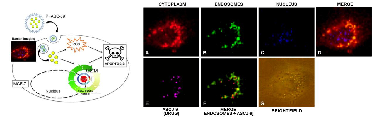
The research on nanoencapsulated drugs for targeted therapies is rapidly emerging. On the other hand, the study of the mechanisms which regulate the intracellular delivery of nanoencapsulated drugs and the cellular distribution of these nanoformulations after their internalization into the cell are still not very clear and difficult to study. Most of the time a fluorescent drug-mimetic or a fluorescent-tagged polymer, have to be used instead of the original ones in order to follow the itinerary of the encapsulate drug.
By using Raman spectroscopy we already demonstrate the possibility to monitor the intracellular delivery of the original drug encapsulated into the original polymer without the use of additional antibodies and with an elevated subcellular resolution. In the figures below we show MCF7 (breast cancer) cultured cells treated with a new anticancer molecule encapsulated into PLGA nanoparticles. In this study we efficiently demonstrated the presence of the drug (magenta) in the perinuclear region, with a partial colocalization with endosomes and related intracellular vesicles (green).
Label-free Raman based assessment of different pathological and physiological cellular types or states.
The diagnosis of several diseases, including cancer, is based on morphological aspects that still nowadays are assessed by visual examination (for example by the evaluation of blood / bone marrow smears). This approach is therefore subjective and scarcely accurate.
On the contrary, due to the chemical specificity of Raman spectroscopy, it is possible to distinguish different cells on the basis of the Raman fingerprint which characterize different cells and is therefore possible to reach elevated diagnostic accuracy. In addition, these methods are potentially be coupled to automatic procedures increasing efficiency and objectivity of the diagnostic procedure.

In the figure above, we show cancer cells derived from patients affected by different acute myeloid leukemia subtypes. Other than have obtained Raman images representative of the morphology of these different leukemia subtypes (middle) , the method was able to automatically distinguish these different cells on the basis of their Raman fingerprint obtained measuring the overall Raman spectra of each cell (right).
Reference: Vanna R., P. Ronchi, Lenferink A.T.M., Tresoldi C., Morasso C., Mehn D., Bedoni M., Picciolini S., Terstappen L., Ciceri F., Otto C. and Gramatica F.. “Label-free imaging and identification of typical cells of acute myeloid leukaemia and myelodysplastic syndrome by Raman microspectroscopy”. Analyst, 2014, Accepted Manuscript. DOI: 10.1039/C4AN02127D