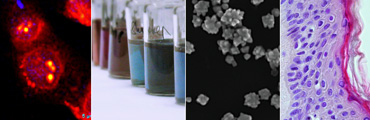

Nanostructured solid surfaces excel with several amazing features in the recent photonics research that are promising to be translated into biomedical applications. They are superior over their flat surface counterparts not only regarding their high surface area, altered hydrophobicity or mechanical behavior, but also regarding their optical properties. Nanostructured noble-metal surfaces fabricated by bottom-up or top-down methods are excellent candidates as substrates for Surface Enahanced Raman, Surface Enhanced Fluorescence, MALDI or Surafce Plasmon Resonance applications. Besides of this, these materials still profit on the high stability and facile phase separation in heterogeneous reaction systems and can be 2D patterned with capture molecules offering the possibility of multiplexing in microarray based bioanalytical assays.
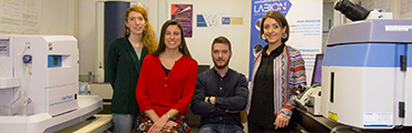
Using in house manufactured materials or in collaboration with industrial (Plasmore S.r.l) and academical (CNR-IMEM Parma, UNIMIB) partners our group is working on development, characterization, and functionalization of nanostructured solid surfaces. Several possible candidates have been characterized during the last three years at Labion by Scanning Electron Microscopy and regarding Raman enhancing properties. The development of a DNA microarray based on solid SERS substrates is currently ongoing as part of the InNaSERSS project (Euronanomed II).
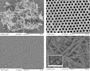
Form top down, left to right:Scanning electron micrographs of ZnO tetrapods (CNR-IMEM, parma) , gold coated polystyrene beads (Plasmore), polymeric nanopillar-gold array (Plasmore), paper based SERS substrate coated with gold nanoparticles.
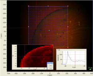
SERS intensity mapping of a spot on a nanopillar-gold SERS substrate