
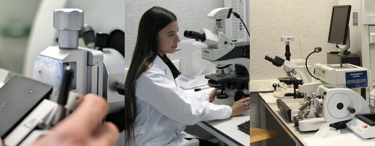
The new LABION Laboratories accommodate several instruments and equipment required for responding to several diagnostic and research issues by using different approaches.
Indeed, the laboratories are equipped with both traditional methods used in chemistry, biochemistry, molecular biology and histology, together with more innovative biophotonic instruments like the Raman microscopes.
Equipments
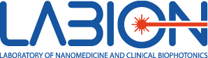
Raman spectrometer
Raman spectroscopy is a non destructive vibrational spectroscopy able to provide the chemical composition of the analyzed sample by a simple irradiation with laser light. Differently from IR, Raman spectroscopy is not affected by the presence of water and therefore is particularly attractive for biomedical studies.
Aramis (Horiba Jobin-Yvon) is a state of the art confocal micro-Raman spectrometer fully controlled through Labspec 6 software with advanced imaging functionalities. The system is equipped with three different laser lines operating at at 532 nm (500mW), 633 nm (50 mW) and 785 nm (500 mW) and has three interchangeable gratings with 600 l/mm 1200 l/mm, 1800 l/mm. The maximum spectral resolution is of about 1 cm-1 wavenumber using the 1800 l/mm grating.
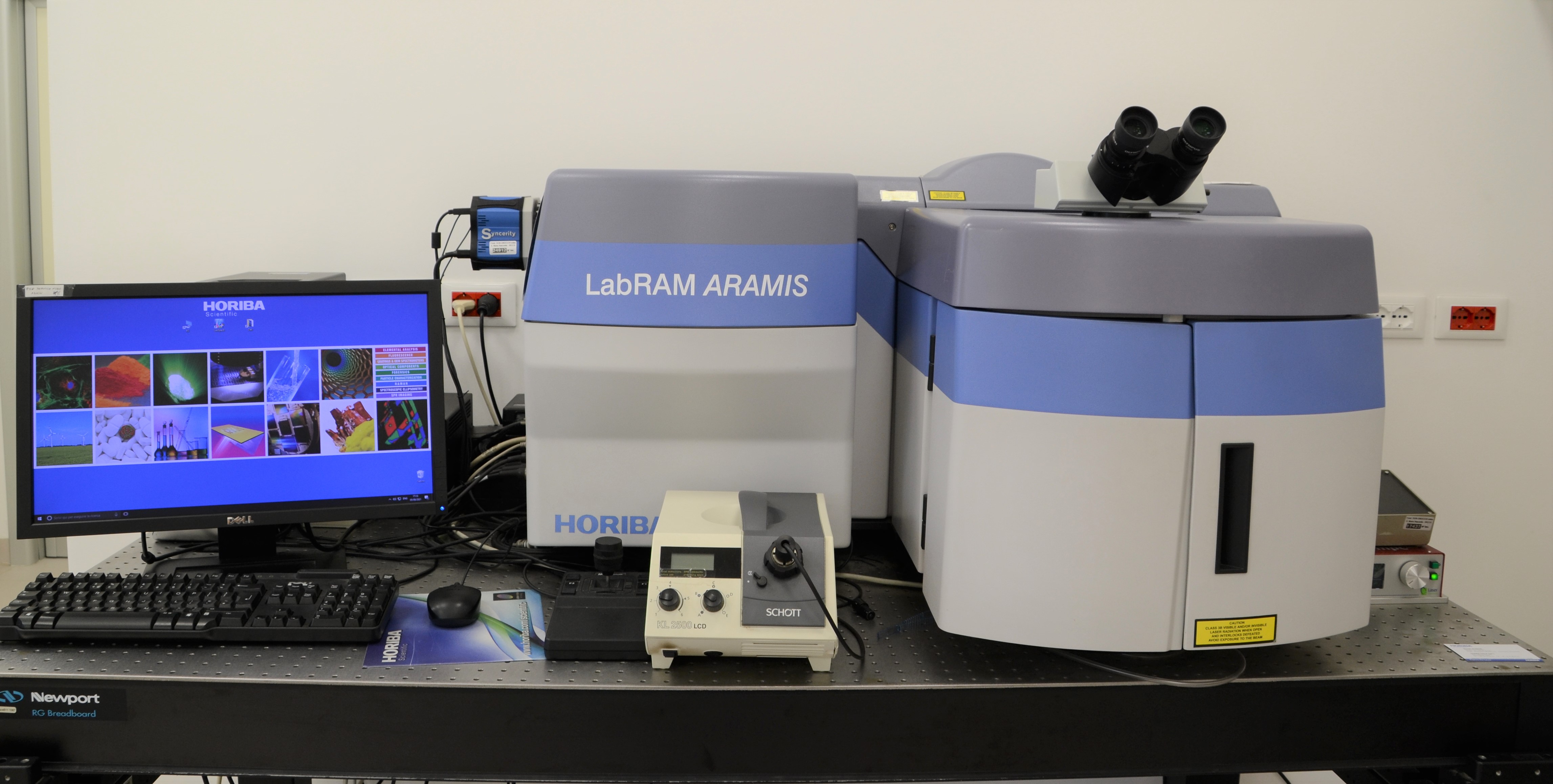
Surface Plasmon Resonance imaging
LABION is the first world wide user of XelPlex (Horiba Jobin-Yvon), a new high-performance and full-automated instrument based on Surface Plasmon Resonance imaging (SPRi). SPRi is based on the direct and label-free detection of the interaction between two molecules (e.g. proteins, smallpeptides, antibodies, drugs, DNA, vesicles, bacteria). The first molecule (“ligand”) is normally immobilized on the gold surface of the SPRi-chip and can then capture the molecule of interest (“analyte”) contained in the sample (e.g. blood, serum, urine). Taking advantage of small changes in the refractive index of the gold surface illuminated by a laser, the SPRi can directly measure the interaction between the two molecules. EzPlex allows the concomitant and fast multiplex detection of hundreds of different analytes in multiple samples and biologic fluids to monitor simultaneously and efficiently several biomarkers related to the disease status, the efficacy of therapeutic treatments and the effect of rehabilitation.
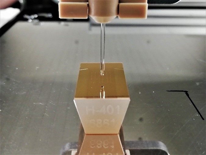
iFOUR Dispensing system
iFOUR Dispensing System of M24You is an automatic instrument for handling of small amounts of chemical or biological solutions or suspensions. In particular, in our laboratory this tool is used to accurately dispense pico/nanoliter drops of ligands onto SPRi custom-specific substrates with high precision and frequency, thanks to a Piezo Driven Micro-Dispenser (PDMD) equipped with a 130 mm long borosilicate glass capillary and a cylindrical piezo ceramic actuator bonded to it.
The instrument includes the software inDot with which it is possible to coordinate the PDMD and create the microarray of interest. Furthermore, iFOUR system allows to collect images of spots, thanks to a camera positioned near the PDMD, in order to visualize the shape and the positions of the spotted ligands.
https://www.m24you.com/surface-plasmon-resonance-imaging/
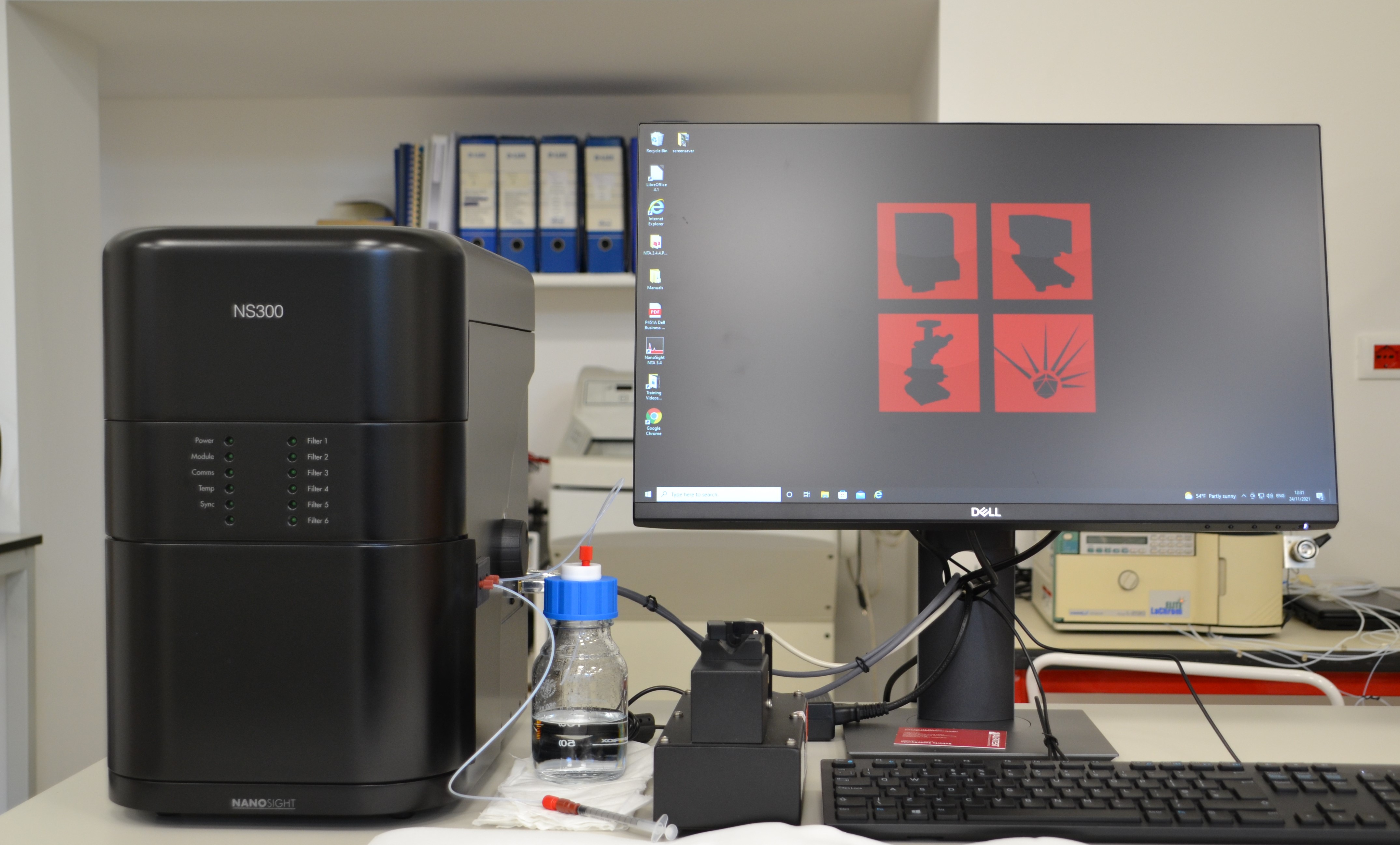
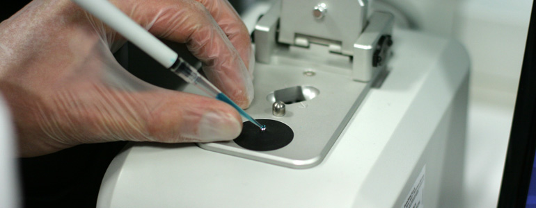
Nanoparticle Tracking Analysis (NTA)
NanoSight NS300 uses Nanoparticle Tracking Analysis (NTA) technology, that utilizes the properties of light scattering and Brownian motion to derive the concentration and size of nano-sized particles. The sample is inserted into the instrument with a syringe and is subsequently hit by a laser. The particles suspended in the path of this laser beam diffuse the light and, consequently, can be viewed thanks to a microscope with 20X magnification on which a camera is mounted. With NanoSight it is also possible to work in fluorescence: by marking the particles with a fluorophore we are able to observe the emission of fluorescence relative to the single particle. More specifically, we use this tool for the characterization of extracellular vesicles, liposomes and metal nanoparticles.
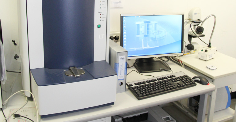
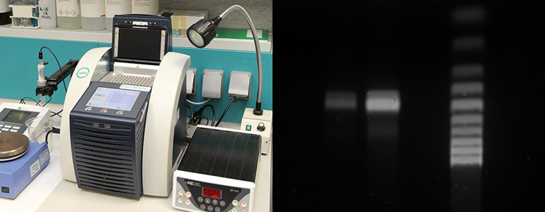
Mass spectroscopy (MALDI-TOF)
The MALDI-TOF (Bruker Daltonics Microflex LT) is a bench-top mass spectrometer used for the analysis of proteins, other small molecules (lipids, carbohydrates) and microorganism (bacteria). Thanks to this instrument is possible to perform fast and reliable analysis for biomarker discovery and for diagnostic purposes. In addition to the high mass accuracy and sensitivity, one of the most relevant advantages of this instrument is the easy, fast and reproducible sample preparation.
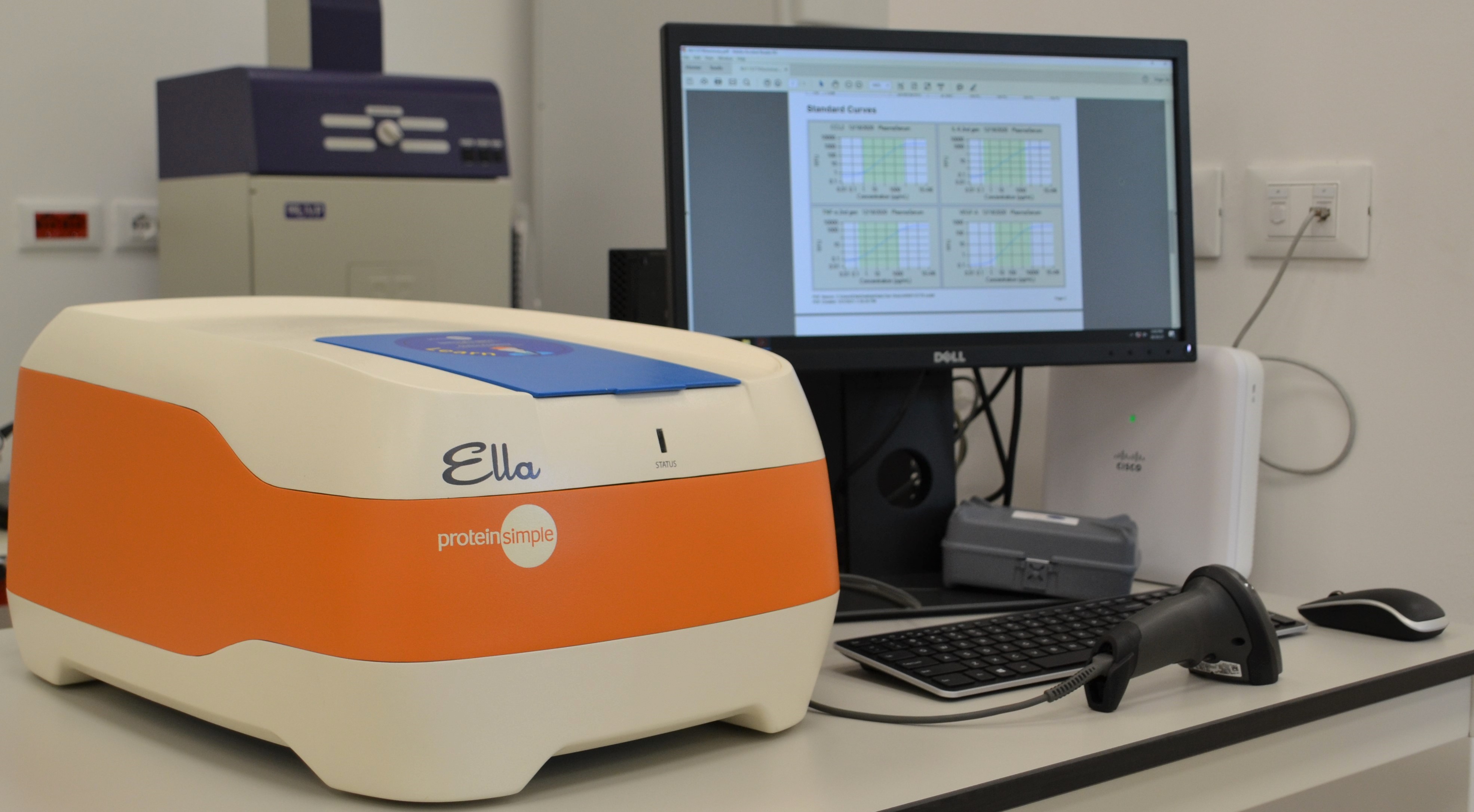
ELLA
Ella is an instrument that allows automated ELISA assays where everything is pre-loaded onto a specific cartridge. After having added sample and buffers and loaded the cartridge into the instrument, in just 90 minutes Ella gives us highly reproducible validated assay data with no manual steps. The assay performance behind that data includes sub-picogram level sensitivity, with high reproducibility. Ella is used in the lab for the characterization of biological fluids and for the detection of markers present in extracellular vesicles samples.
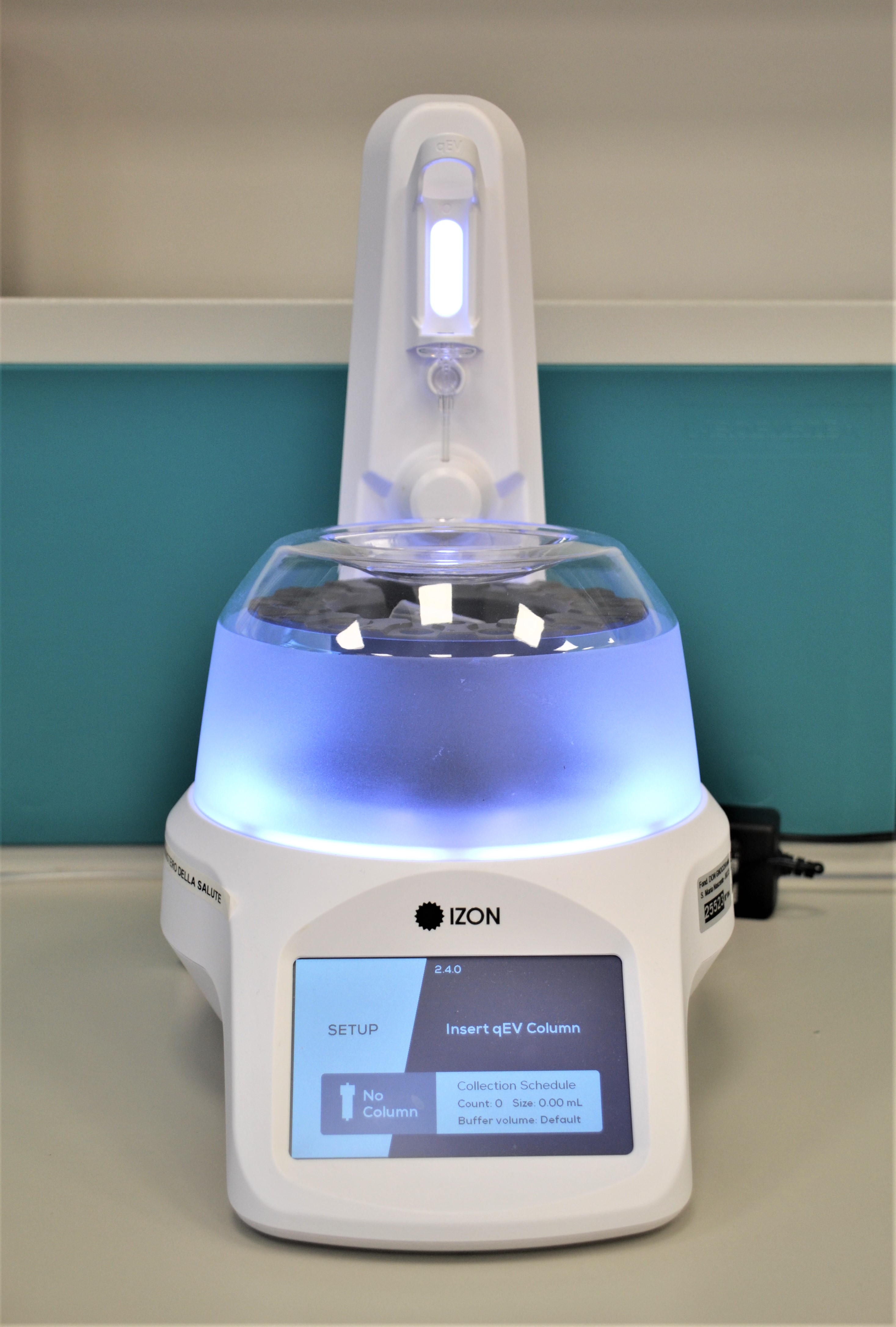
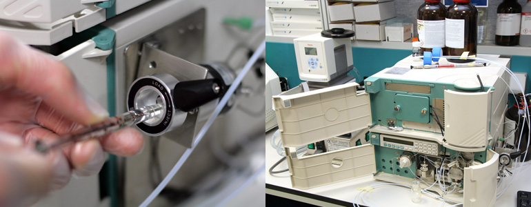
AUTOMATIC FRACTION COLLECTOR (AFC)
Izon Automatic Fraction Collector (AFC) allows for fast, precise, automated isolation of extracellular vesicles by size-exclusion chromatography using IZON columns. It utilizes a rotational carousel for holding collection tubes where fractions of extracellular vesicles are collected. Once thebuffer volume has been collected, the instrument begins collecting a predetermined number of fractions of a given volume as set by the user. During fraction collection the carousel advances to the next fraction when the selected fraction weight is reached. It is a compact instrument ideal for small scale and high throughput parallel processing of sample, such as blood, urine and saliva.
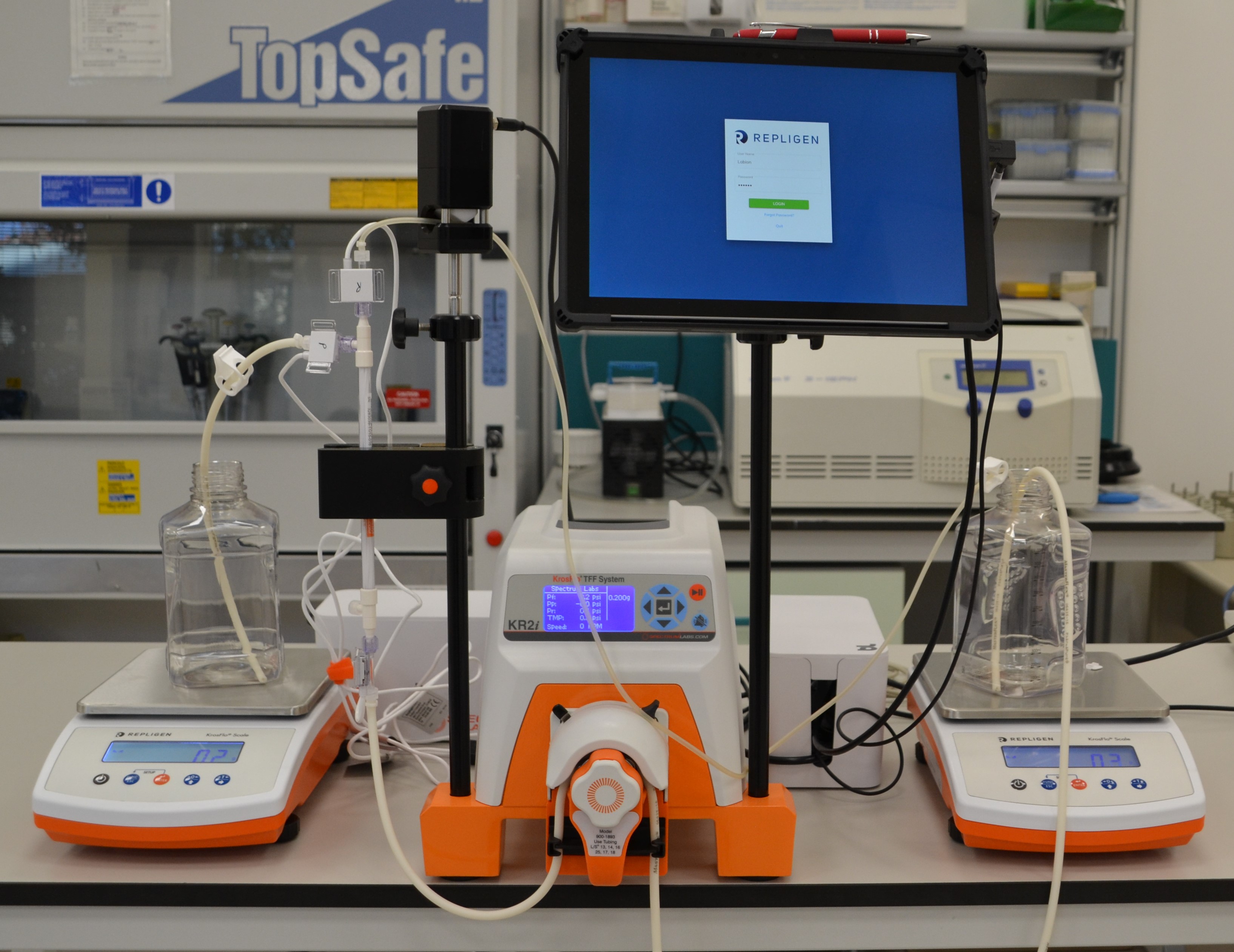
TANGENTIAL FLOW FILTRATION
Tangential Flow Filtration (TFF) is a particular process of separation, different from other filtration systems. Indeed, the fluid is passed parallel to the filter rather than being pushed through a membrane perpendicularly, which can clog the filter medium. The particles that pass through the membrane form the permeate, while the particles that are retained form the retentate. This tool allows to concentrate a molecule of interest within a solution and, furthermore, it allows the separation of small and large particles (diafiltration). In particular, we use TFF to isolate extracellular vesicles from different suspensions.
PCR and related molecular biology techniques
The PCR is the universal technology used for the amplification of a genetic sequence (DNA) derived from both real clinical samples or from synthetic processes. This method can be used for several research and clinical purposes, in particular, in our laboratory PCR is used for the production of oligonucleotides modified in order to be coupled to nanostructured components and as golden standard technique for comparing and validating the performances of innovative biophotonics/nanotechnology-based methods developed in our laboratory.
- DNA Extraction
- RNA Extraction
- cDNA production
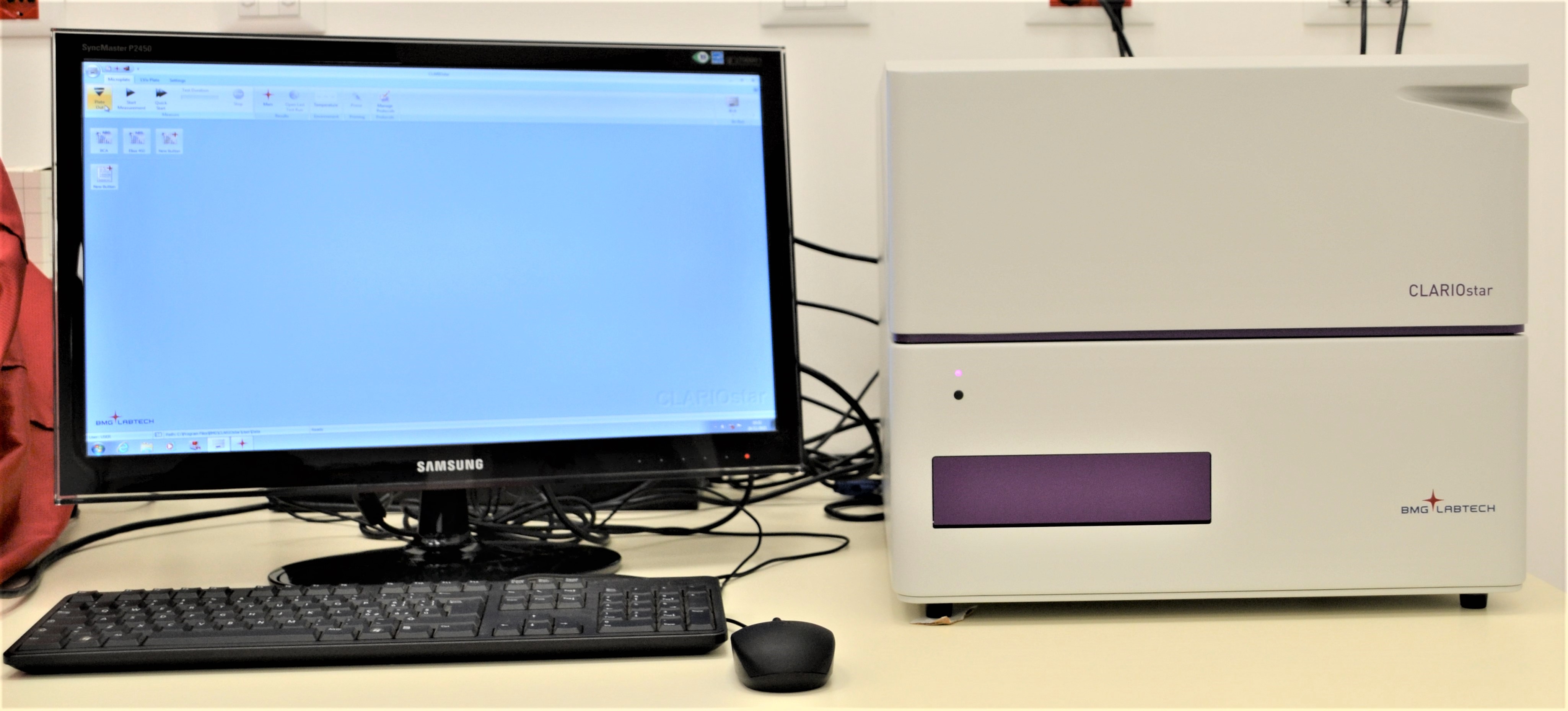
Microplaste reader
The microplate reader CLARIOstar (BMG LABTECH) is a high performance multimodal detection system, equipped with a combination of monochromators, filters, and spectrometer that make it suitable for a variety of applications in the different detection modes, like for example molecule quantization, kinetic study of chemical, enzymatic and biological reactions, ELISA assays.
It is suitable for the multiplexing analysis of mutiwell until 1536 wells with high speed acquisition of the absorbance in the UV/vis spectrum (220 – 1000 nm with 1 nm of resolution).
CLARIOstar dual monochromator technology provides high sensitivity in fluorescence studies with a precise selection of excitation and emission wavelengths, reducing the background signal.
UV/VIS Spectrophotometer (Nanodrop)
The NanoDrop 2000 is a wide spectral range (190-840 nm), easy to use UV-Vis Spectrophotometer for a big variety of applications including analysis of peptides, DNA, RNA, protein, nanoparticle samples, colorimetric assays, dyes. Works with normal and micro plastic and quartz cuvettes or using the nanodrop function requires only 0.5 – 2 µL of the sample. The user friendly software includes pre-configured methods for DNA, Protein A280, Microarray, Protein and Labels, Pierce 660, Bradford, BCA, and Lowry and has various data export capabilities.
HPLC
High-performance liquid chromatography (HPLC) is a typical multipurpose analytical method for the separation and identification of several class of molecules. This instrument can be used for both the identification of biomarkers in clinical samples (e.g form blood, serum, urine, tissue homogenate) and for the identification of compounds derived from chemical reactions or cells cultured in-vitro.
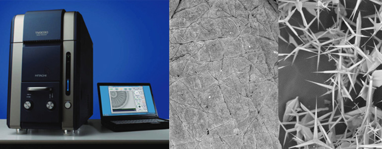
Scanning electron microscope (SEM)
The Hitachi TM3000 table top SEM enables taking good resolution images without special sample preparation in a very short time. The low vacuum mode allows imaging of hydrated or non-conducting samples and two selectable beam energies (acceleration voltage) help imaging of soft or hard matter. A high-quality multi-segment electron detector provides contrast-rich images with selectable material/compositional or topographical emphasis. Thanks to the simplicity of the software, users can fast become independent operators after short training. The large stage allows introduction of vide variety of samples related to life science, food, cosmetics, healthcare, pharmaceutical, textiles or materials science.
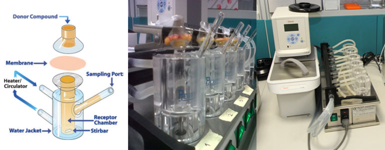
Franz-type diffusion cell
The Franz Cell is a simple, reproducible test for in-vitro skin permeation studies. Franz-type system is relatively simple in the design (see the scheme below), the cell comprises two parts: the donor chamber that contains the substance (e.g. drug , nanoparticles) to be tested and the receptor chamber containing the medium with the substance diffused and the sampling port. The two parts are separated by a membrane, human skin in our experiments. The receptor temperature is usually maintain to 32 degrees C°, to approximate normal skin conditions, thanks to a heater circulator.
The samples are collected for several hours and analysed using HPLC or similar technique.
LABION is equipped with a complete system of 6 vertical Franz-type diffusion cells (Permegear), stirrer station and thermostated water machine (see the figures below).
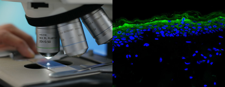
Histology instruments and services
Histological analysis is one of the most widespread techniques in life science research, useful both for diagnostic purposes and basic biomedical studies. Chemical fixation and paraffin embedding of tissue samples allow long lasting preservation of the physiological morphology and the molecular composition of the biopsy. Taking advantage of a manual rotary microtome, it is possible to obtain serial thin paraffin sections (2-10 µm) suitable for subsequent histological staining and immunolocalisation techniques. Routine staining procedures guarantee the observation of the overall tissue cytoarchitecture, whereas structural protein expression might be highlighted by means of multistep immunological reactions.
In our laboratory, frozen samples can be cryosectioned thanks to a cryostat designed to prevent abrupt temperature variation of the sample while sectioning. High resolution, bright field images are obtained with an upright light microscope equipped with digital camera.
Fluorescence Microscope
The Leica DM2500 upright microscope is designed for the observation of histological sections in bright field, differential interference contrast, polarization contrast, and high-performance fluorescence. Chromatically-corrected optical paths ensure high contrast images from 25x to 630x magnification. For epi-fluorescence microscopy, thanks to three integrated filter blocks, it is possible to select filters and dichroic mirrors according to the fluorophore used, thus obtaining the optimal fluorescence signal with minimal background light. The camera associated software with a multi-channel imaging capability allows instant high resolution image overlay, morphometric analysis, and image processing.
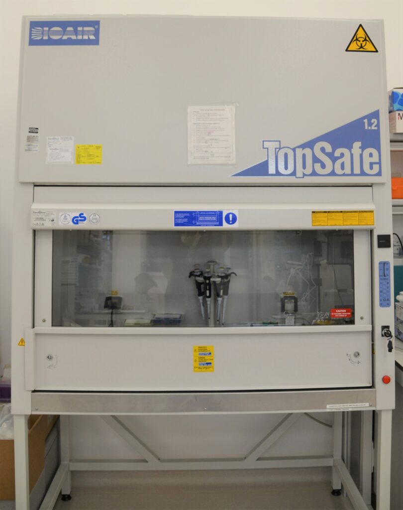
Biological safety cabinet
The biological safety cabinet ensures the aseptic or particulate free conditions needed for tissue and cell culture experiments. It uses filters and directional horizontal laminar air flow to provide a contaminant-free work area. The horizontal flow hood belongs to the Class II biological safety cabinets which protect the specimen, the user, and the environment from contamination and it is suitable for applications requiring sterile conditions without the need to work with volatile chemicals.
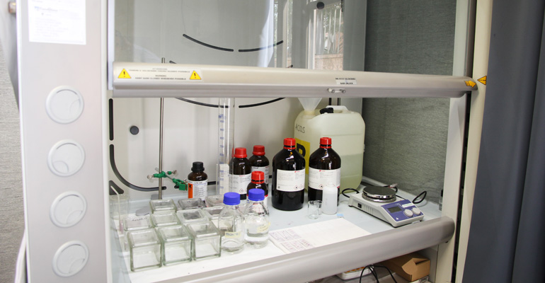
Laboratory chemical hood
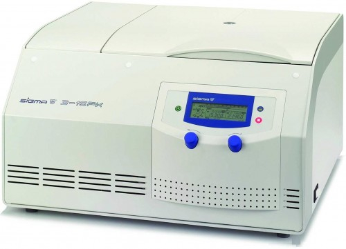
Refrigerated Centrifuge SIGMA 3-16PK